A Shining Light for Cataract Patients

(above) Michael V. Drake, MD, President of the University of California with Daniel Schwartz, MD

Infographic about donation impact with QR code.

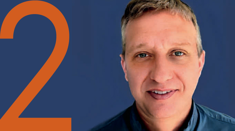
This past spring, a remarkable breakthrough in cataract technology brought renewed hope to patients seeking improved vision. All May See’s President Emerita, Kathleen Rydar, underwent cataract surgery at UCSF, where Associate Professor Julie Schallhorn, MD, MS, performed the procedure using an innovative Light Adjustable Lens™ (LAL). These groundbreaking lenses, developed by UCSF’s very own Daniel Schwartz, MD, in collaboration with Nobel Laureate, the late Robert Grubbs, PhD, and Julia Kornfield, PhD, from Caltech, revolutionize the post-surgery experience for patients by fine-tuning the visual correction using only ultraviolet light.

The origins of the LAL date back 25 years when Dr. Schwartz first envisioned the concept. As a retinal specialist, he was inspired to create an intraocular lens made of a light-sensitive material that could be adjusted non-invasively using a laser after the eye had healed and the refractive errorwas stable. This idea sparked a fruitful collaboration with Dr. Grubbs, leading to the realization of this groundbreaking technology. The U.S. Food and Drug Administration (FDA) approved the LAL in 2017, offering patients who had undergone cataract surgery the possibility of optimized vision without relying on glasses.

Close-up of a woman with light hair in a dark jacket.
Kathleen Rydar, All May See’s President Emerita

The All May See Foundation played a crucial role in supporting the initial efforts of the collaboration at Caltech, fostering the development of this extraordinary technology.
 

President Emerita receives light adjustable lenses

One of the first recipients of the LAL at UCSF, Ms. Rydar attests to its effectiveness, expressing her delight at the outcome. After three adjustment sessions, she can now read effortlessly and enjoy the beauty of the world, even spotting wildflowers on mountaintops without the aid of glasses.

A woman in a white coat smiling beside ophthalmology equipment.
Associate Professor, Julie Schallhorn, MD, MS

Dr. Schallhorn, who performed the surgery, emphasizes the significant impact the LAL has had on her surgical practice. While existing techniques for selecting intraocular lens power are highly effective, some patients still require glasses. With the light adjustable lens, the risk of such errors is substantially decreased. Patients can now experience clear vision without their cataract and fine-tune their eyesight to their exact preferences, marking a momentous advancement in post-cataract surgical care.

Dr. Schallhorn is enthusiastic about the future of ophthalmology, particularly with the continuous development of new technologies. Reflecting on the field’s progress over the past 50 years, she marvels at how cataract surgery has transformed from a procedure with significant risk of potential vision loss to a routine
outpatient surgery.

Dr. Schallhorn and her colleagues at UCSF, alongside other visionaries in the field, are committed to exploring new ways to treat vision impairment and continue pushing the boundaries
of ophthalmic innovation.

Text about Light Adjustable Lens innovations.

The Light Adjustable LensTM represents a shining beacon of hope for cataract patients, illuminating a future where clearer vision and improved quality of life are within reach for countless individuals around the world.


Lap for Landon: Extending UCSF Ophthalmology’s Impact Beyond the Bay Area

A family with a stroller at an outdoor event.
Brian Woods (dad), Courtney Woods (mom), Everett Woods (brother), and Landon Woods (in stroller)

FAMILY INITIATIVES

The impact of UCSF Ophthalmology reaches far beyond the Bay Area, as exemplified by a heartwarming tribute to Landon, a young boy diagnosed with Gould Syndrome, organized by his family from St. Louis, Missouri. Despite the pouring rain on October 30, 2022, more than 150 adults and children gathered to celebrate Landon’s first birthday through a Lap for Landon walk to raise awareness for his condition.

One-third of infants diagnosed with Gould Syndrome have cataracts or developmental defects leading to early-onset glaucoma. This rare, multisystem disorder named after Douglas Gould, PhD, Professor and Vice President for Research for UCSF Ophthalmology, honors his groundbreaking discovery and tireless research in this area.

The syndrome results from mutations in collagen genes COL4A1 and COL4A2 responsible for the support and reinforcement of body tissues.

The family’s effort contributed over $12,000 from the walk to support Dr. Gould’s vision research, aiming to make a lasting difference in treating this uncommon ailment.

Spreading the word proves fruitful

All May See has raised awareness and funds for Gould Syndrome for more than a year. Our journey began on Rare Disease Day 2022 with a generous $125,000 challenge gift from a Texas family whose 9-year-old son had recently been diagnosed with Gould Syndrome (featured in Vision Summer 2022). On Rare Disease Day and Gould Syndrome Day 2023, we issued a second challenge to reach $250,000 in donations. Through the kind contributions of individuals and families worldwide affected by Gould Syndrome, we have raised over $187,000.

The fund’s purpose is to gain a deeper understanding of the underlying mechanisms of Gould Syndrome. Dr. Gould’s lab is actively exploring therapeutic avenues, including the potential use of CRISPR gene-editing technology, which holds promise.

The St. Louis family is holding the Second Lap for Landon on Saturday, October 21st. To support this effort in honor of Landon and individuals affected world-wide, please visit the All May See Foundation donation page (allmaysee.org/donate) and select “Lap for Landon 2023” from the drop-down menu under “Designation.”

Two-Year Update on All May See Research Awards

Every year, All May See awards grants to Ophthalmology researchers to facilitate preliminary studies focused on novel approaches to discover the causes and treatments for a wide range of vision problems. In 2021, a total of $200,000 in award grants were provided for the following projects:

 
A man in glasses smiling with a green background.Project Title: National Eye Institute (NEI) Center Core Grant for Vision Research
Principal Investigator: Erik Ullian, PhD

Findings: The acquisition of a state-of-the-art Zeiss LSM900 Airyscan 2 confocal microscope was made possible with NEI funds and a generous gift from Don and Judy McCubbin. This microscope is crucial for the success of numerous NEI-funded projects. To ensure its optimal performance, the All May See grant funds the cost of two additional years of service contract to maintain the microscope in excellent condition.


A man smiling with a blue background and a large orange number two.Project Title: Elucidating Mechanisms of Visual Pathway Damage in Alzheimer’s Disease
Principal Investigator: Alexander Smith, PhD

Findings: The research discovered that amyloid beta peptides associated with Alzheimer’s Disease accumulate in the optic nerve and the sclera. These findings offer a novel mechanistic explanation for vision loss in Alzheimer’s disease. Dr. Smith intends to seek National Institutes of Health (NIH) funding to further this research in collaboration with the National Institute on Aging.


Portrait of a woman smiling with glasses against a blue background with an orange number 3.
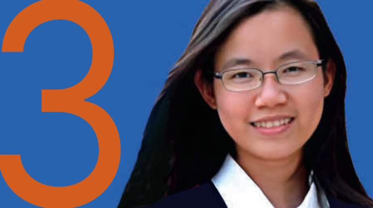
Project Title: Mechanisms of Retinal Degeneration in Alzheimer’s Disease-Related Dementias
Principal Investigators: Li Xuan Tan, PhD (Postdoctoral Fellow in the laboratory of Aparna Lakkaraju, PhD)

Findings: Dr. Tan’s investigation shed light on the mechanisms responsible for impaired visual function in frontotemporal dementia (FTD). The research discovered specific defects in energy production and metabolism in the tissue that nourishes and protects the retina, leading to inflammation and loss of the light-sensing cells of the eye, resulting in progressive visual deficits.


Smiling man with short hair and a gray shirt beside a large orange number four.Project Title: Enabling Direct Correlation of Choroidal Blood Flow and Retinal Degeneration at the Single Vessel Level and Over Time Using Transscleral Multiphoton Microscopy
Principal Investigator: Henk Shang, PhD (Postdoctoral Fellow in the laboratory of Tyson Kim, MD, PhD)

Findings: The research team made significant progress in developing transscleral imaging and analytical methods to study changes in blood vessels within a genetic model of retinitis pigmentosa. Their findings revealed significant shrinking of blood vessels and diminished blood flow in diseased eyes, prompting further investigation into the correlation between these changes and the progression of retinal degeneration.


A smiling woman with long, wavy hair against a light background, with the number 5 displayed prominently in orange.
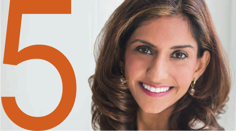
Project Title: Transcutaneous Orbicularis Oculi Stimulation for Temporary Eyelid Closure
Principal Investigators: Nailyn Rasool, MD (pictured) and Julie Schallhorn, MD, MS (not pictured here)

Findings: The principal investigators (PI‘s) successfully developed a prototype device in partnership with the Prototyping Lab at the Cleveland Veteran’s Administration Hospital. This prototype effectively induced temporary lid closure. The ability to induce lid closure will be extremely effective in patients with neurodegenerative conditions preventing lid closure as well as in patients with neurotrophic keratopathy. The goal is to prevent the development of thinning and ulceration of the cornea without permanent closure of the eyelid. The PI’s have also filed for intellectual property protection for this device design. Additionally, they established a connection with Israeli company Neurotrigger, who are working on a similar device to act as a blink pacer for patients with Bell’s palsy. Dr. Schallhorn joined their team as a consultant and aided in the development of plans to bring their device to the US, pending FDA approval.

New Research Awards

At their April 2023 meeting, the Board of Directors of All May See approved $200,742 in research awards for the following projects:


Orange number one on a white background.Development of an injectable biopolymer device for sustained intravitreal delivery of carbonic anhydrase inhibitor for the treatment of juvenile X-linked retinoschisis.


Principal Investigator: Robert Bhisitkul, MD, PhD

A man in a suit and glasses smiling at the camera.
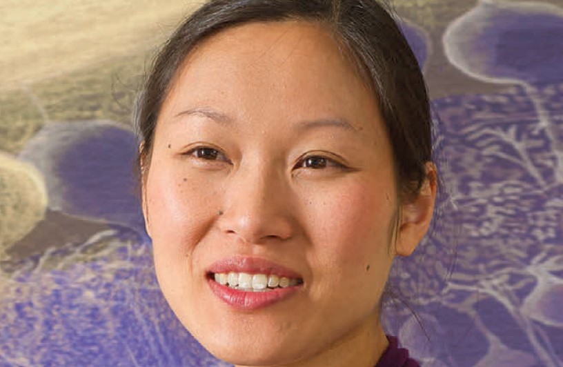
Juvenile X-linked retinoschisis (XLRS) is an inherited retinal disorder (IRD) that predominantly affects boys and men, arising in early childhood with potential blindness by their teens or adulthood. While there are few medication options for IRDs, XLRS responds well to carbonic anhydrase inhibitor (CAI) treatment. Treatment with eye drops has been effective but is unable to fully penetrate the eye and dependent on patient application. This project aims to develop a minimally invasive injectable device to provide CAI therapy for 6–12 months, offering a better treatment approach.


Large orange number two on a white background.
Identification of a visually evoked reflex as a more sensitive diagnostic for graded cone loss.


Two women smiling in separate portraits.

Principal Investigators: Felice Dunn, PhD, and Jacque Duncan, MD

Many degenerative vision diseases are caused by the deterioration of photoreceptors (light-sensitive cells) in the back of the eye. However, this loss can evade detection until more than half of the cells are compromised. The Dunn and Duncan labs are collaborating to develop high-sensitivity assays to detect photoreceptor degeneration earlier than current methods. The Dunn lab’s discoveries revealed novel changes to visual processing following modest cell loss, and in combination with the clinical expertise of the Duncan lab in diagnosing such conditions, this project hopes to generate a new window into eye health, enabling the early identification, management, and mitigation of degenerative vision diseases.


The number three in bold orange font.Deep learning system to predict 5-year risk of high myopia using fundus imaging in a North American population of children.


A young man in glasses wearing a suit, smiling.

Principal Investigator: Jonathan Li, MD

This project involves photographing North American children’s retinas and processing the photos through an artificial intelligence system to predict a child’s risk of developing severe nearsightedness (high myopia). If found valid, this non-invasive approach will enable the identification of patients most in need of aggressive treatment while there is still ample time for intervention.


Large orange number four on white background.Multiphoton Aqueous Flowmetry and Image-Guided Laser Therapy: Novel Approaches for Glaucoma Precision Medicine.


A scientist in a lab coat using a pipette.

Principal Investigator: Tyson Kim, MD, PhD

Currently, the only treatment to slow or stop progression of glaucoma is to lower eye pressure. This project investigates the use of advanced laser imaging techniques for new, less-invasive methods to measure the drainage of fluid (aqueous humor) from the eye, which will ultimately result in more precise methods of glaucoma evaluation and treatment.

More than meets the eye

Unveiling the Risk of Diabetic Eye Disease Progression
 
A woman smiling in a blue blazer.
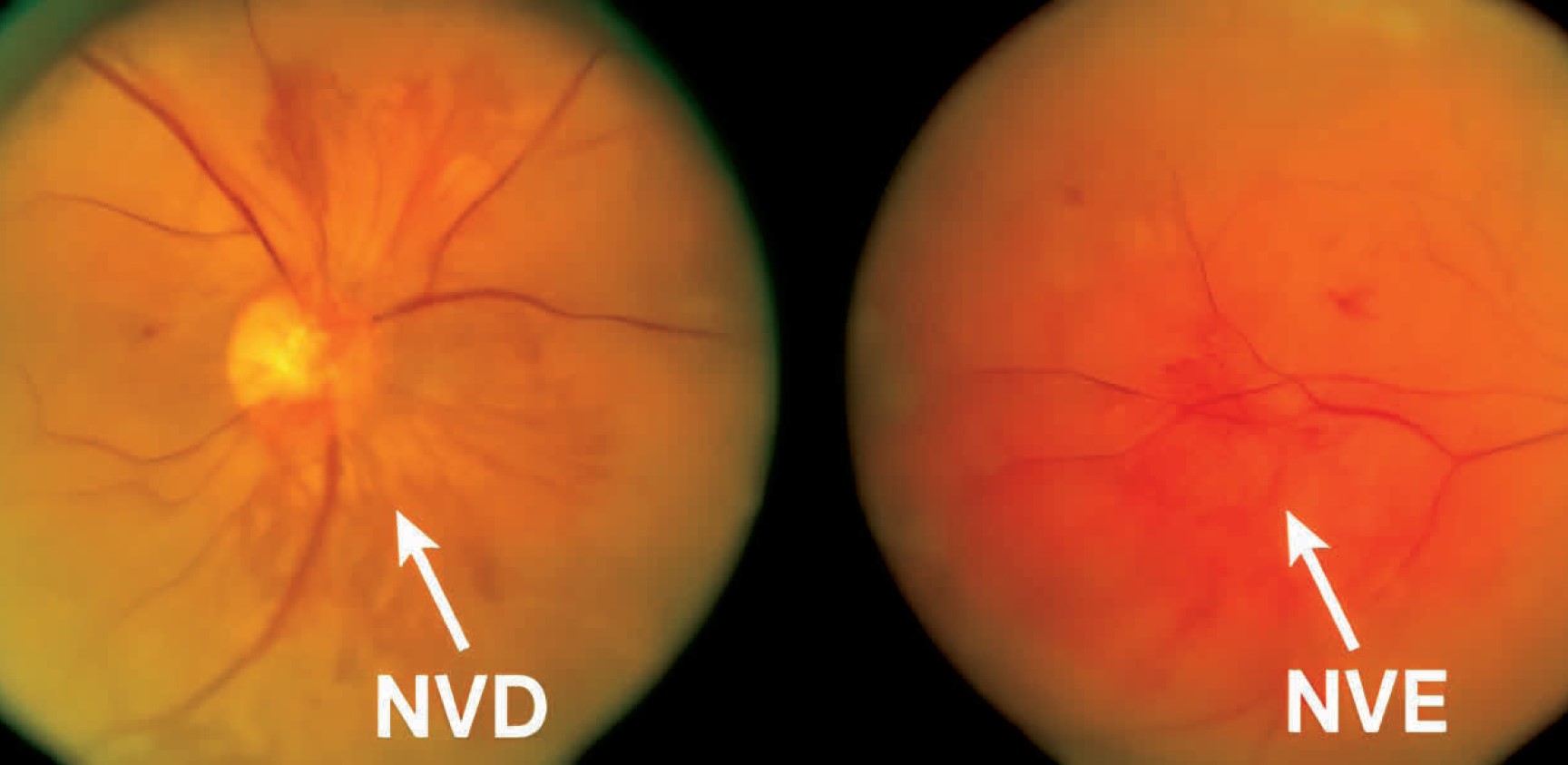
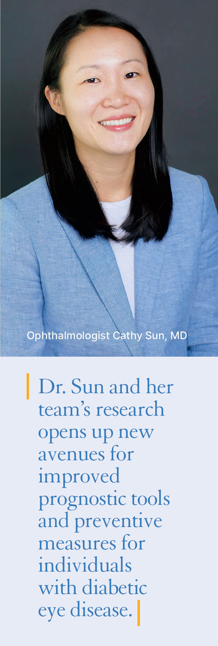
Diabetic retinopathy is a critical eye disease that poses a significant threat to vision if left untreated. To address the issue of inadequate preventive care and the absence of a reliable method to predict high-risk patients, ophthalmologist Cathy Sun, MD, and her team at UCSF embarked on a groundbreaking study. Their aim was to develop predictive models for the progression of diabetic retinopathy, enabling healthcare providers to implement timely interventions and prevent vision loss in affected individuals.
 

Analyzing influencing factors

Drawing from patient medical records at UCSF Health and Zuckerberg San Francisco General (ZSFG) Hospital, the research team meticulously analyzed various influencing factors that could predict the advancement to more severe stages of the disease. Key factors such as insurance coverage, age, and average Hemoglobin A1c levels, which provide insights into blood sugar control, emerged as critical risk factors significantly affecting the progression to more severe diabetic retinopathy.
 

Personalized risk assessment tool

The study’s findings paved the way for the development of a personalized risk assessment tool that goes beyond general population-level predictions. Dr. Sun and her team aspire to create a tool capable of predicting the risk of progression on an individual patient level. By leveraging the patient’s complete medical information, this tool would empower healthcare providers to make more accurate assessments of individual risk. Consequently, timely interventions could be implemented to halt disease progression and successfully prevent vision loss.
 

Improving patient outcomes

The study conducted by Dr. Sun and her team at UCSF sheds light on critical risk factors for diabetic retinopathy progression. Their endeavor to develop a personalized risk assessment tool demonstrates a commitment to improving patient outcomes. As this research progresses, healthcare providers may gain a valuable ally in the fight against diabetic eye disease, enabling them to implement timely and effective interventions that preserve the precious gift of sight for countless individuals.

Two eye fundus images showing NVD and NVE.
These are examples of an eye with proliferative diabetic retinopathy (PDR) with neovascularization of the optic disc (NVD) and neovascularization elsewhere on the retina (NVE).

Copenhagen Award & Members-in-Training Outstanding Poster Award Winner

Nicole Tsai


Copenhagen Award 2022

We are pleased to announce that Nicole Tsai from Dr. Xin Duan’s lab has won the David and Joyce Copenhagen Prize for Best Published Paper in Visual Sciences for papers published in 2022. Nicole is being recognized for her Nature Neuroscience paper describing the development of Trans-Seq to identify retinotectal synapses.

This award was established to recognize a UCSF graduate student, postdoctoral fellow or clinical fellow mentored by a faculty member in the Department of Ophthalmology who has published outstanding work in the area of laboratory visual sciences.
 
Read the paper here: https://www.nature.com/articles/s41593-022-01068-8
 

Members-in-Training Outstanding Poster Awards at ARVO 2023

Nicole also recently won the MIT Outstanding Poster Award at this year’s ARVO meeting in the BI Section.

MIT First Authors of the top five (5) scored abstracts scheduled as Poster presentations from each Scientific Section and Cross-sectional Group were eligible and invited to participate in the MIT Outstanding Poster Award Competition held at the ARVO Annual Meeting.

 
Our warmest congratulations to Nicole for her achievements!