

The UCSF Department of Ophthalmology congratulates Matilda F. Chan, MD, PhD, Professor in the Department of Ophthalmology and member of the Francis I. Proctor Foundation, on being selected as a 2026 ARCHES Mid-Career Scholar.
The ARCHES Mid-Career Development Program is a two-year faculty development program that supports UCSF research faculty who are outstanding mentors and have demonstrated a commitment to the PRIDE Values and Principles of Community. Scholars receive protected research time, mentorship, and career development opportunities to help advance their research and leadership.
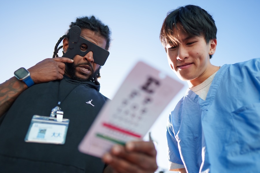
Dr. Chan’s research focuses on the cellular and molecular mechanisms involved in corneal repair and corneal endothelial dystrophies. She also studies barriers to ophthalmic care, with a particular interest in patients experiencing housing insecurity.
Dr. Chan is also active in scientific publishing. She serves on the editorial boards of three vision journals and is Managing Director of the Council of Vision Editors Fellowship (CVEF). The fellowship provides mentored training in peer review and editorial practice for early-career ophthalmology and optometry faculty.
Dr. Chan is one of ten faculty members selected for the 2026 ARCHES Mid-Career Scholar cohort.
Learn more about the 2026 ARCHES Mid-Career Scholar cohort, or visit Dr. Chan’s UCSF Profile Page to learn more about her research and clinical interests.


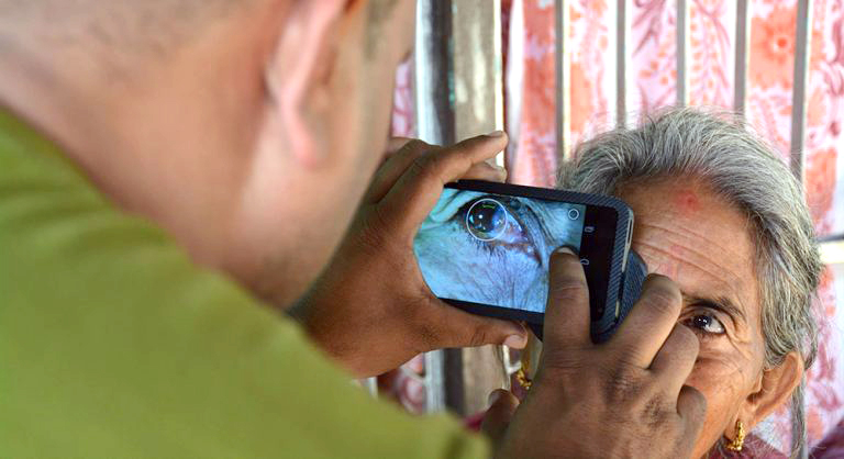

 Jeremy Keenan, MD, MPH, is the H. Bruce Ostler Professor of Ophthalmology and the Director of International Programs at the Proctor Foundation. He is an epidemiologist and ophthalmologist with clinical specialties of cornea and uveitis. His research focuses on strategies to reduce the global burden of blindness, with an emphasis on developing countries.