A recent UCSF News story highlights the UCSF eye clinic at the Division Circle Navigation Center. A student-run initiative founded in 2015, the clinic brings vision care directly to people experiencing homelessness in San Francisco.
Each month, UCSF medical students, ophthalmology residents and fellows, faculty ophthalmologists and optometrists, and community partners at St. Vincent de Paul come together to provide eye exams, connect patients to follow-up care, and replace lost or stolen glasses. The clinic serves as both a vital community resource and a hands-on learning opportunity for the next generation of eye care providers.
The article offers a powerful snapshot of the clinic in action and the impact vision care can have on patients’ lives. As the story notes, for some patients, “new glasses can mean a driver’s license, a chance to land a job, housing, and even schooling.” The clinic also gives learners the opportunity to engage directly with patients in the community while developing clinical skills under the mentorship of residents, fellows, faculty, and optometrists.
Read the full story to learn more about the dedicated students, trainees, faculty, staff, and community partners who make this work possible and help restore patients’ independence and quality of life.who make this work possible and help restore patients’ independence and quality of life.
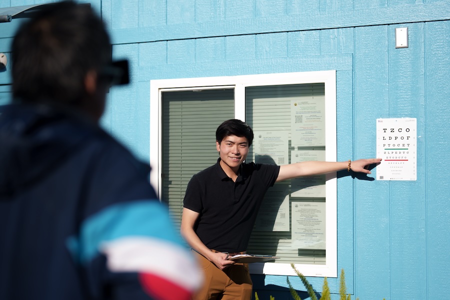

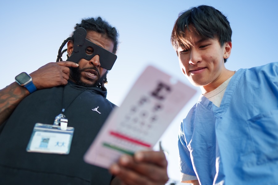
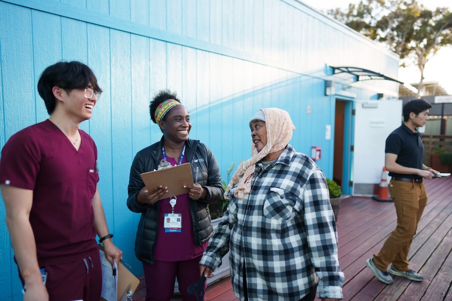

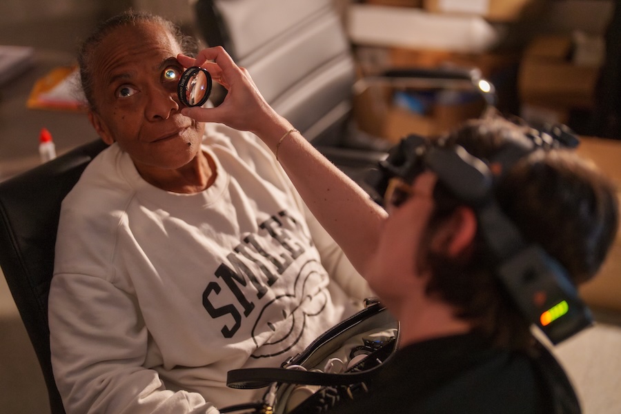

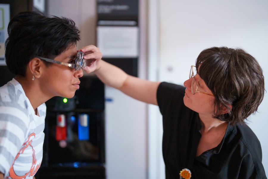
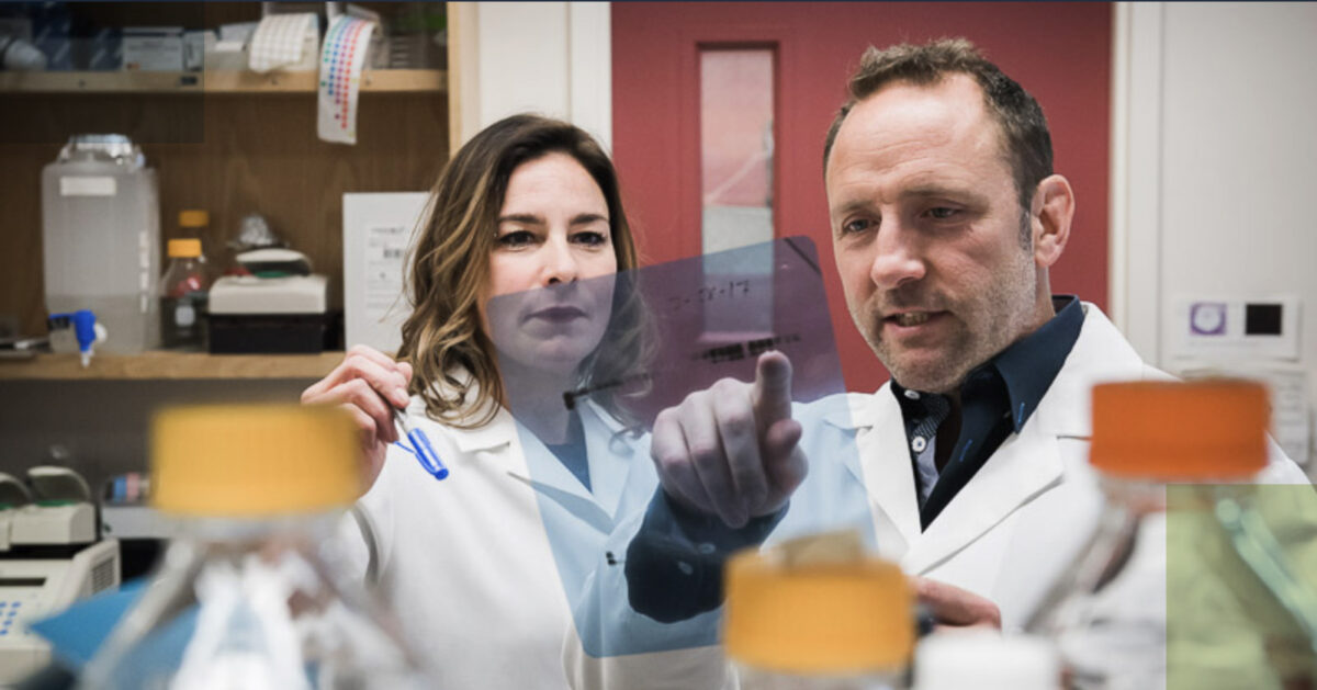
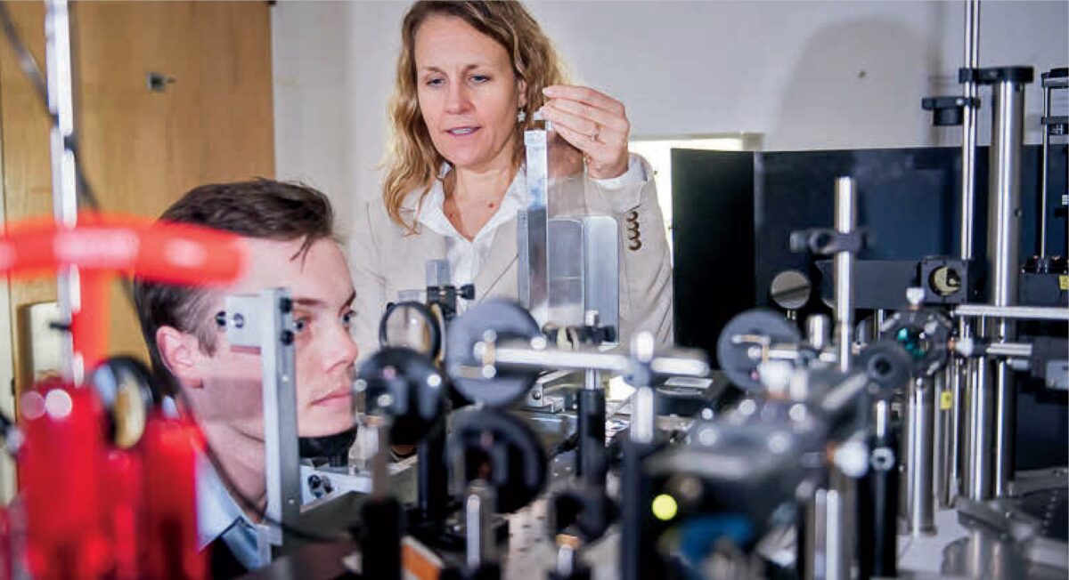
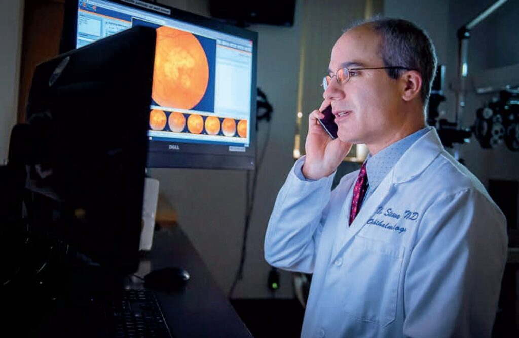
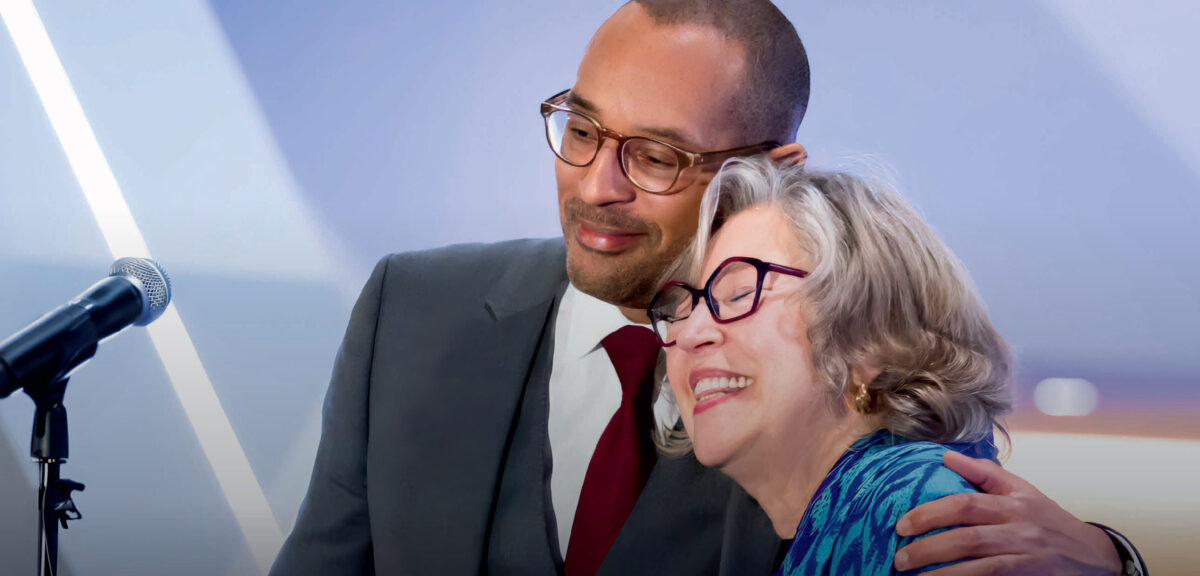
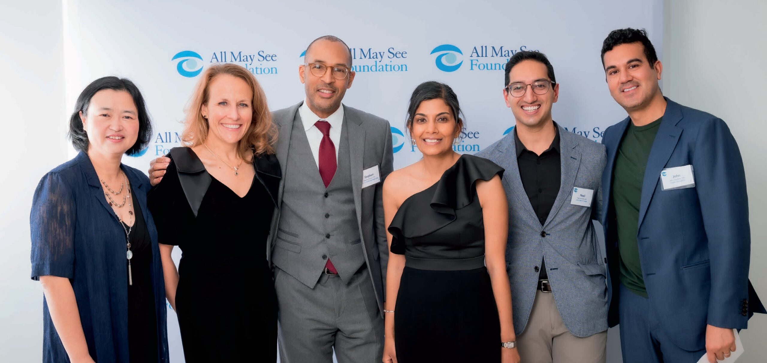
Scenes from a recent community outreach clinic, UCSF Ophthalmology Shelter Clinic, a team of medical students and residents providing free comprehensive eye exams and vision care to homeless patients at the Division Circle Navigation Center (a St. Vincent de Paul-operated shelter in San Francisco SoMa district), on May 13, 2026. Photos by Maurice Ramirez.
Read the full UCSF News story: A Clinic That Helps San Francisco Eyes See Clearly