FAMILY INITIATIVES
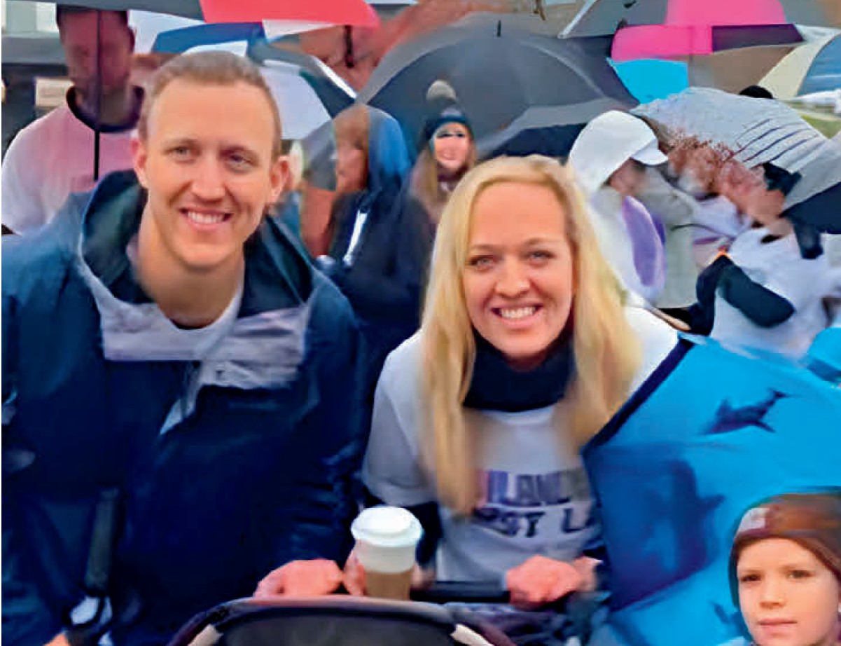
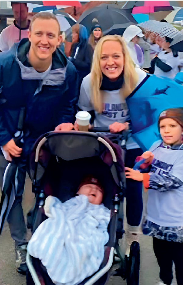
The impact of UCSF Ophthalmology reaches far beyond the Bay Area, as exemplified by a heartwarming tribute to Landon, a young boy diagnosed with Gould Syndrome, organized by his family from St. Louis, Missouri. Despite the pouring rain on October 30, 2022, more than 150 adults and children gathered to celebrate Landon’s first birthday through a Lap for Landon walk to raise awareness for his condition.
One-third of infants diagnosed with Gould Syndrome have cataracts or developmental defects leading to early-onset glaucoma. This rare, multisystem disorder named after Douglas Gould, PhD, Professor and Vice President for Research for UCSF Ophthalmology, honors his groundbreaking discovery and tireless research in this area.
The syndrome results from mutations in collagen genes COL4A1 and COL4A2 responsible for the support and reinforcement of body tissues.
The family’s effort contributed over $12,000 from the walk to support Dr. Gould’s vision research, aiming to make a lasting difference in treating this uncommon ailment.
Spreading the word proves fruitful
All May See has raised awareness and funds for Gould Syndrome for more than a year. Our journey began on Rare Disease Day 2022 with a generous $125,000 challenge gift from a Texas family whose 9-year-old son had recently been diagnosed with Gould Syndrome (featured in Vision Summer 2022). On Rare Disease Day and Gould Syndrome Day 2023, we issued a second challenge to reach $250,000 in donations. Through the kind contributions of individuals and families worldwide affected by Gould Syndrome, we have raised over $187,000.
The fund’s purpose is to gain a deeper understanding of the underlying mechanisms of Gould Syndrome. Dr. Gould’s lab is actively exploring therapeutic avenues, including the potential use of CRISPR gene-editing technology, which holds promise.
The St. Louis family is holding the Second Lap for Landon on Saturday, October 21st. To support this effort in honor of Landon and individuals affected world-wide, please visit the All May See Foundation donation page (allmaysee.org/donate) and select “Lap for Landon 2023” from the drop-down menu under “Designation.”
 Project Title: Elucidating Mechanisms of Visual Pathway Damage in Alzheimer’s Disease
 Project Title: Mechanisms of Retinal Degeneration in Alzheimer’s Disease-Related Dementias
 Project Title: Enabling Direct Correlation of Choroidal Blood Flow and Retinal Degeneration at the Single Vessel Level and Over Time Using Transscleral Multiphoton Microscopy
 Project Title: Transcutaneous Orbicularis Oculi Stimulation for Temporary Eyelid Closure
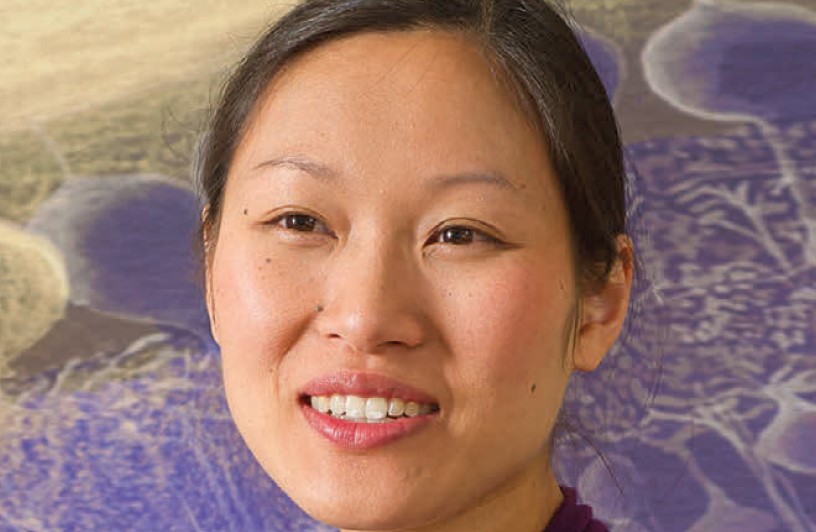
 Juvenile X-linked retinoschisis (XLRS) is an inherited retinal disorder (IRD) that predominantly affects boys and men, arising in early childhood with potential blindness by their teens or adulthood. While there are few medication options for IRDs, XLRS responds well to carbonic anhydrase inhibitor (CAI) treatment. Treatment with eye drops has been effective but is unable to fully penetrate the eye and dependent on patient application. This project aims to develop a minimally invasive injectable device to provide CAI therapy for 6–12 months, offering a better treatment approach.
 Multiphoton Aqueous Flowmetry and Image-Guided Laser Therapy: Novel Approaches for Glaucoma Precision Medicine.
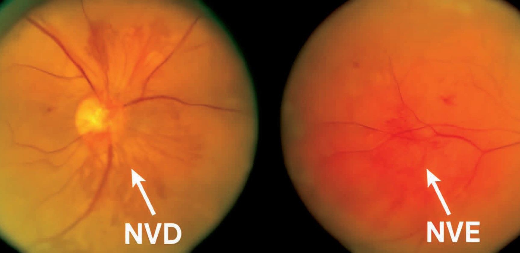
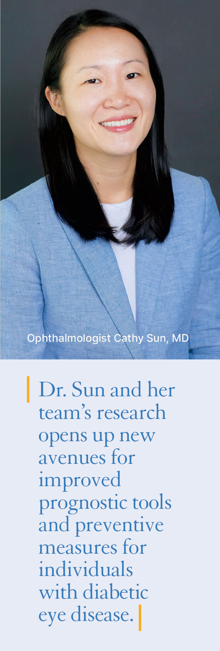 Diabetic retinopathy is a critical eye disease that poses a significant threat to vision if left untreated. To address the issue of inadequate preventive care and the absence of a reliable method to predict high-risk patients, ophthalmologist Cathy Sun, MD, and her team at UCSF embarked on a groundbreaking study. Their aim was to develop predictive models for the progression of diabetic retinopathy, enabling healthcare providers to implement timely interventions and prevent vision loss in affected individuals.