Ying Han, MD, PhD

Professor


Novel Microfluidic Meshworks for Use in Glaucoma Drainage Implants

Dr. Han, Professor of Ophthalmology, Co-Director of the Glaucoma service, and Glaucoma Fellowship Director, is a glaucoma specialist whose research addresses the need for improved and fibrosis-free surgical implants for glaucoma. In collaboration with bioengineers and material sciences, she leads NIH-funded work developing novel microfluidic meshworks for use in glaucoma drainage implants. In addition, she has organized a multicenter international RCT to examine the role of intra- and postoperative mitomycin-C for use with Ahmed glaucoma valves. Other projects include integrated wireless microactuators for fibrosis control in glaucoma drainage devices and microfluidic contact lens sensors for intraocular pressure monitoring.


To Learn More:

https://profiles.ucsf.edu/ying.han


Research Areas:

Glaucoma
 
Learn more about UCSF Ophthalmology faculty research.

Bob Bhisitkul, MD, PhD

Professor


Age-Related Macular Degeneration Treatment and New Retinal Therapies

Dr. Bhisitkul, Professor of Ophthalmology and Director of the Retina Fellowship, is a vitreoretinal specialist who in collaboration with UCSF’s bioengineering group has developed a novel thin-film drug delivery system that can produce long term zero order kinetics through regulating pore sizes at nanoscale. This is now being tested <em>in vivo</em> for delivery of anti-VEGF agents. In addition to his bioengineering projects, he directed the SEVEN-UP cohort study of long-term outcomes in age-related macular degeneration treatment and is an investigator in major clinical trials of new retinal therapies. As Director of the Retina Fellowship, Dr. Bhisitkul has mentored many clinical fellows as well as research fellows. Scholars interested in ocular drug delivery and clinical trials will be interested in the Bhisitkul lab.


To Learn More:

https://profiles.ucsf.edu/bob.bhisitkul


Research Areas:

Macular Degeneration, Retina or Retinal Diseases
 
 
Learn more about UCSF Ophthalmology faculty research.
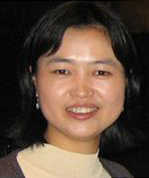
Erik Ullian, PhD

Professor


Synapse Formation and Function in the Visual System

Dr. Ullian, Professor of Ophthalmology, studies molecular mechanisms that regulate synapse formation and function in the visual system. He has used cell culture systems to screen for genes and small RNAs that impact synaptic transmission. His laboratory has identified the miRNA pathway as an important regulator of a variety of neuronal and synaptic processes relevant to visual system development. The Ullian lab is also studying the interactions between neurons and glia that are required for proper development and function of the nervous system. He is developing astrocyte, neuron, and microglial 3D organoids to model key aspects of neurodegenerative diseases. In addition to graduate students and postdoctoral fellows, Dr. Ullian has mentored K awardees, including Dr. Yvonne Ou. Scholars who are interested in the interactions of neurons and glia in the visual system will be interested in the Ullian lab.


To Learn More:

https://profiles.ucsf.edu/erik.ullian
https://ullianlab.ucsf.edu/


Research Areas:

Visual System Development, Retina or Retinal Diseases, Stem Cell Research
 
Learn more about UCSF Ophthalmology faculty research.
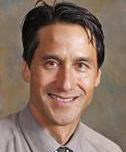
Yvonne Ou, MD

Associate Professor


Cellular and Synaptic Mechanisms of Glaucomatous Neurodegeneration

The Ou laboratory studies the cellular and synaptic mechanisms of glaucomatous neurodegeneration. We are particularly interested in understanding the earliest steps of inner retina circuit disassembly in response to injury. Our research program combines imaging and analysis of specific cell and synaptic labels in the retina, rodent models of experimental glaucoma, novel genetic tools in which specific cell types are labeled, and the tools of molecular/cell biology and physiology to address a series of questions that, unanswered, have prevented progress in the field: 1) What are the early steps of compartmentalized neurodegeneration of the ganglion cell in glaucoma? 2) Are there specific ganglion cell types that are more susceptible to intraocular pressure (IOP) elevation? 3) Are there specific inner retinal circuits that are more susceptible to IOP elevation? As a clinician-scientist, I am motivated to advance our field not only by answering these fundamental questions, but also by translating the knowledge gained into improvements in diagnostic and treatment modalities in glaucoma. For example, a detailed understanding of the earliest structural and functional changes that occur in glaucoma will allow us to design treatments that can rescue RGCs, perhaps the most susceptible RGCs, before irreversible cell death occurs.


To Learn More:

https://profiles.ucsf.edu/yvonne.ou
https://ophthalmology.ucsf.edu/oulab/


Research Areas:

Glaucoma, Mobile Health, Visual System Function in Adults
 
 
Learn more about UCSF Ophthalmology faculty research.

Kayarat Nair, PhD

Assistant Professor


Mechanisms of Glaucoma and Ocular Growth

The Nair lab studies the genetics and biology of ocular diseases with a primary focus on glaucoma and relevant phenotypes. Glaucoma is a heterogeneous group of diseases characterized by death of retinal ganglion cells, specific visual field deficits, and optic nerve degeneration. It is a leading cause of blindness worldwide and affects over 70 million people. The genes and molecular mechanisms contributing to glaucoma are poorly understood. Our goal is to identify genes, molecular mechanisms and cellular networks contributing to glaucoma. Individuals with refractive errors (both myopia or hyperopia) are at a higher risk of developing glaucoma. Another focus of the lab is to understand the role of refractive error in the pathogenesis of glaucoma. Our laboratory employs a variety of multi-disciplinary approaches to dissect the role of complex disease associated genes at a mechanistic level. We have established important mouse models that recapitulate features of the human disease. They provide us the platform to discover new genes/pathways, elucidate the molecular mechanisms, contributing to glaucoma as well as to test hypothesis and new treatment ideas. In parallel, we employ tools of human genetics as a path towards the identification of disease genes.


To Learn More:

https://profiles.ucsf.edu/kayarat.nair
https://ophthalmology.ucsf.edu/nairlab/


Research Areas:

Glaucoma, Gene Research, Visual System Development
 
Learn more about UCSF Ophthalmology faculty research.
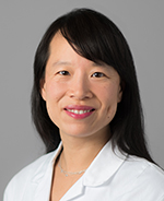
Maxence Nachury, PhD

Professor


The Molecular Basis of Ciliopathies

The Nachury lab studies Bardet-Biedl Syndrome (BBS), a hereditary disease characterized by retinal degeneration, obesity, polydactyly, and polycystic kidneys. The root cause of Bardet-Biedl Syndrome is dysfunction of the primary cilium, a surface-exposed organelle required for vision, olfaction and developmental signaling. Pioneering biochemical and imaging studies by Dr. Nachury’s team have revealed that a complex of BBS proteins traffics signaling receptors out of cilia. The Nachury lab is now identifying the regulation of trafficking out of cilia by BBS proteins. A second research direction is aimed at cilia-derived extracellular vesicles (EVs). Ciliary signaling receptors that fail conventional exit are packaged into ciliary EVs. By dissecting the mechanisms of ciliary EV biogenesis, Nachury and colleagues will gain insight into the roles of these extracellular parcels in cell-cell communication.
 
Research in the Nachury lab is funded by the National Institute of General Medical Sciences (NIGMS), the National Eye Institute (NEI), Research to Prevent Blindness (RPB) and the America Diabetes Association (ADA). Dr. Nachury is the recipient of multiple awards including the 2020 Cogan award from the Association for Research in Vision and Ophthalmology (ARVO).


To Learn More:

https://profiles.ucsf.edu/maxence.nachury
http://nachurylab.ucsf.edu


Research Areas:

Retinitis Pigmentosa or Retinal Degenerations, Cilia, Ciliopathies
 
Learn more about UCSF Ophthalmology faculty research.

Deepak A. Lamba, M.B.B.S., Ph.D.

Associate Professor


Retinal Repair Following Inherited and Age-Associated Degeneration

Dr. Lamba leads an NIH-funded laboratory focused on using human stem cells for retinal repair following inherited and age-associated degeneration. He utilizes stem cell technologies to generate disease-in-a-dish models or for cell replacement. The key research interests include (1) exploring the potential and challenges in retinal cell replacement therapies esp. the role of the tissue microenvironment and (2) developing stem-cell based model systems to understand various human retinal degenerations and identifying new therapeutic avenues including genome editing technologies like CRISPR. Dr. Lamba is a natural collaborator with clinician-scientists and since joining the Department in 2018 he has established collaborations with Drs. Jacque Duncan and Tony Moore. Dr. Lamba has mentored numerous pre- and postdoctorates and is also a member of the education committee. His group will provide opportunities for Scholars interested in harnessing the power of novel stem cell technologies to clinical problems especially retinal degenerations.


To Learn More:

https://profiles.ucsf.edu/deepak.lamba
http://lambalab.ucsf.edu
https://ophthalmology.ucsf.edu/lambalab/


Research Areas:

Gene Therapy, Retinitis Pigmentosa or Retinal Degenerations, Stem Cell Research, Leber Congenital Amaurosis, Retinal Development, Pluripotent stem cells, Cell replacement
 
 
Learn more about UCSF Ophthalmology faculty research.