Kim Lab Research
Novel Technologies for Ocular Imaging and Therapy
Many of the eye’s most important structures sit behind optically opaque tissue and remain difficult to visualize or manipulate in the living eye. Our lab develops imaging, laser-delivery, and computational platforms that overcome these limits, combining advanced multiphoton microscopy, targeted laser strategies, and computer vision-based analysis to resolve previously inaccessible tissues and physiology at the subcellular scale. These platforms enable longitudinal study of the living eye and, paired with genetic, pharmacologic, and biomechanical perturbations, support mechanism-focused investigations that were not previously feasible.
Vascular Development, Physiology, and Aqueous Outflow Regulation
We study how vascular systems form and specialize, with a focus on the relationship between structure, flow, and disease. Our work integrates intravital imaging, transgenic models, and quantitative flow analysis to define the genetic, developmental, and physiologic programs that govern vascular organization, including mechanisms of neurovascular development. We extend these principles to the eye, where we investigate neurovascular function of the retina and how the aqueous outflow pathways regulate fluid drainage. Ongoing efforts aim to define the molecular, cellular, and biomechanical determinants of outflow control in glaucoma and to identify new therapeutic strategies.
Translation, Accessibility, and Clinical Impact
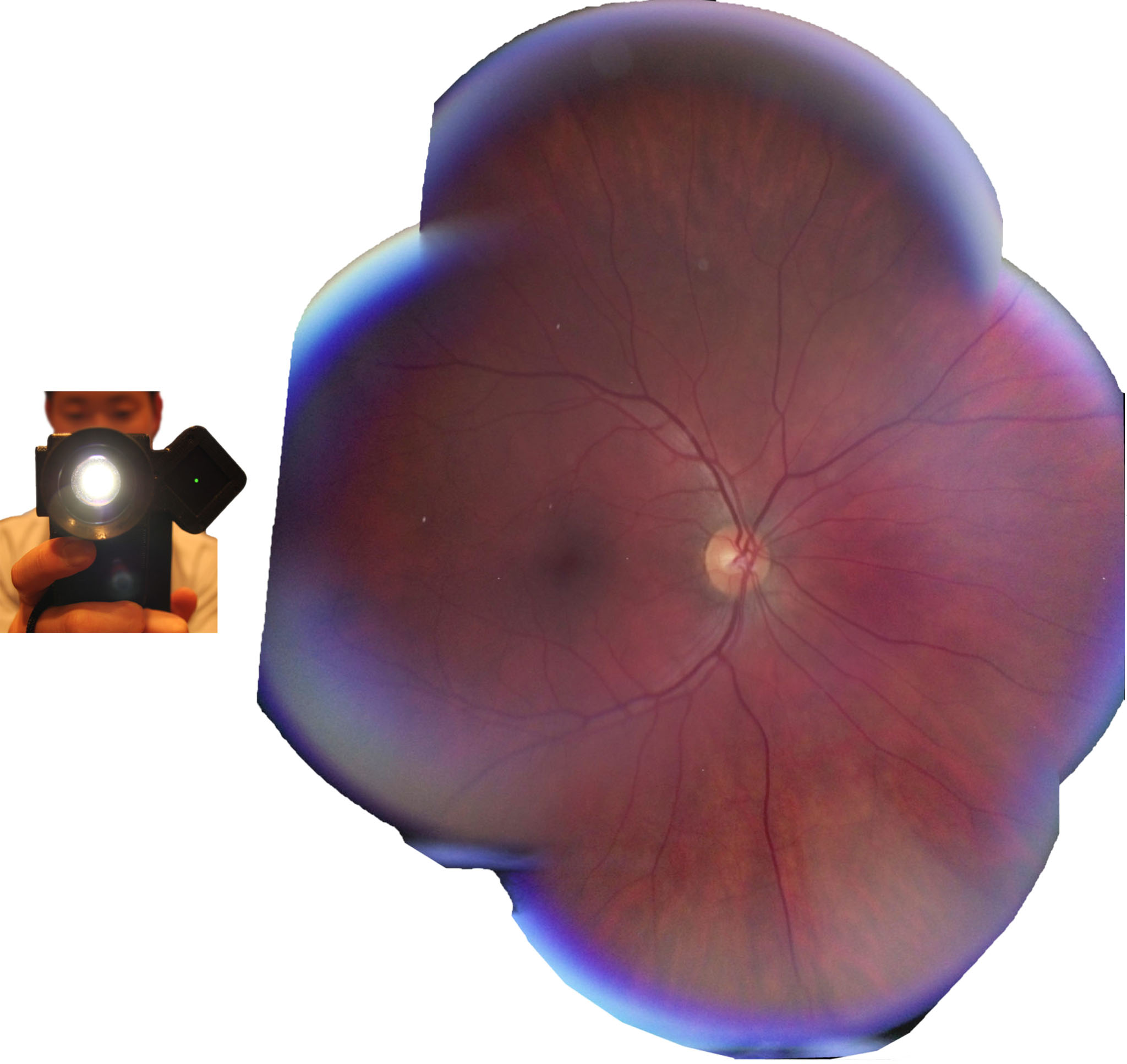
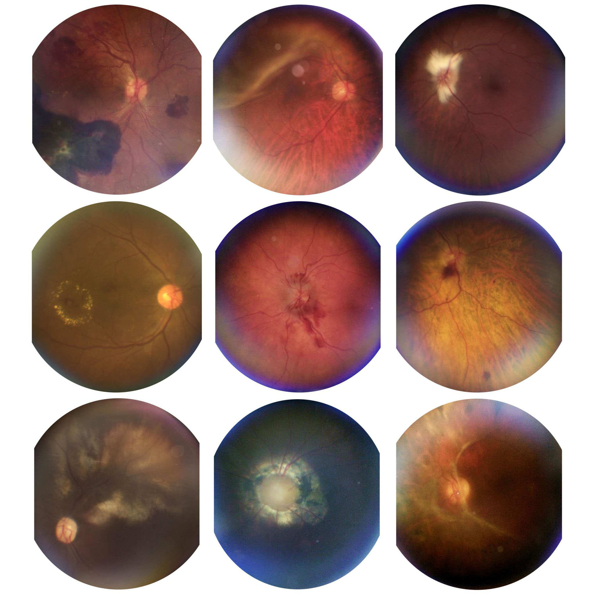
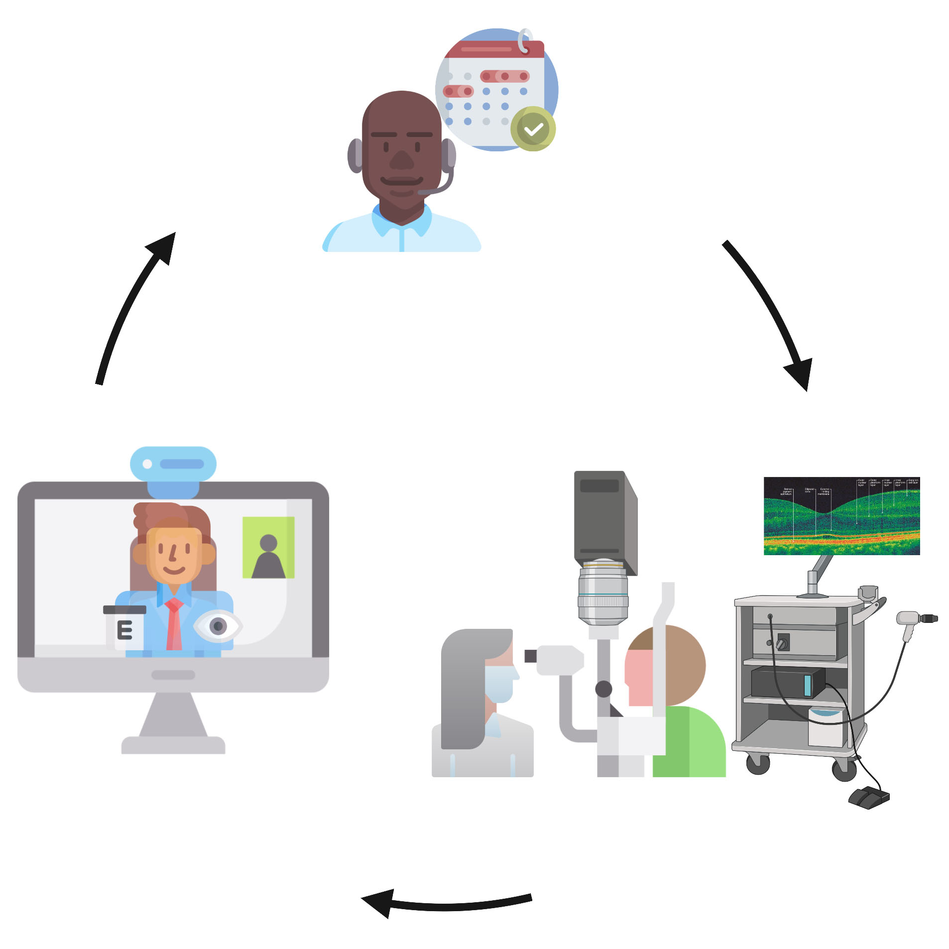
A central goal of the lab is to translate technological innovation into improved patient care, with a particular focus on extending access for underserved populations. We have developed portable, AI-enabled platforms for wide-field retinal screening of diseases such as diabetic retinopathy and are advancing new quantitative assessments of vascular and aqueous physiology in the eye. This work is paired with direct investment in care delivery, including cost-coverage and care-extension programs within safety-net health systems that provide sight-restoring treatments to underserved patients.
Many of the eye’s most important structures sit behind optically opaque tissue and remain difficult to visualize or manipulate in the living eye. Our lab develops imaging, laser-delivery, and computational platforms that overcome these limits, combining advanced multiphoton microscopy, targeted laser strategies, and computer vision-based analysis to resolve previously inaccessible tissues and physiology at the subcellular scale. These platforms enable longitudinal study of the living eye and, paired with genetic, pharmacologic, and biomechanical perturbations, support mechanism-focused investigations that were not previously feasible.
Vascular Development, Physiology, and Aqueous Outflow Regulation
We study how vascular systems form and specialize, with a focus on the relationship between structure, flow, and disease. Our work integrates intravital imaging, transgenic models, and quantitative flow analysis to define the genetic, developmental, and physiologic programs that govern vascular organization, including mechanisms of neurovascular development. We extend these principles to the eye, where we investigate neurovascular function of the retina and how the aqueous outflow pathways regulate fluid drainage. Ongoing efforts aim to define the molecular, cellular, and biomechanical determinants of outflow control in glaucoma and to identify new therapeutic strategies.
Translation, Accessibility, and Clinical Impact
A central goal of the lab is to translate technological innovation into improved patient care, with a particular focus on extending access for underserved populations. We have developed portable, AI-enabled platforms for wide-field retinal screening of diseases such as diabetic retinopathy and are advancing new quantitative assessments of vascular and aqueous physiology in the eye. This work is paired with direct investment in care delivery, including cost-coverage and care-extension programs within safety-net health systems that provide sight-restoring treatments to underserved patients.