Our Research
The Chan lab studies cellular and molecular mechanisms underlying corneal and ocular surface eye diseases. Our team of basic scientists, clinicians, and biostatisticians uses a basic and translational scientific approach to understanding extracellular signals in the corneal microenvironment and identifying biomarkers that are important in vision-threatening diseases including corneal scarring and Fuchs endothelial dystrophy. We aim to apply scientific discoveries to the understanding, prevention, and treatment of ocular diseases.
Learn more about our main areas of research:
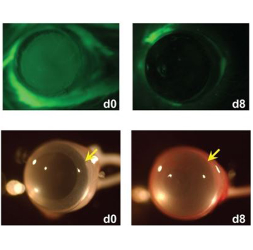
Regulation of Corneal Repair by Metalloproteinases
Metalloproteinases have important regulatory roles in various physiological and pathological processes including inflammation, neovascularization and cell proliferation. We have found that MMP12 (macrophage metalloelastase) is expressed in injured corneas and has a protective role in the inflammatory, neovascular, and fibrotic responses to corneal injury. MMP12 has also been shown by others to have anti-bacterial and anti-viral activities. Using mouse models and human patient samples, we are determining the molecular mechanisms underlying the protective effects of MMP12 after corneal injury and infection.
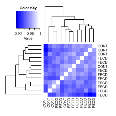
Role of DNA Methylation in Fuchs Endothelial Corneal Dystrophy
Fuchs Endothelial Corneal Dystrophy (FECD) is a progressive, degenerative disease of the corneal layer of the eye, resulting in corneal swelling and vision loss. FECD is a major cause of vision loss and the leading reason for corneal transplantation in older patients. DNA methylation is a basic, cellular process used to control gene function by attaching small chemical tags to DNA. Our lab is comparing DNA methylation changes between normal corneal tissue and diseased corneas obtained from patients with FECD undergoing corneal transplantation.
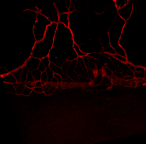
Multivalent Anti-VEGF Conjugates for Sustained Treatment of Diabetic Retinopathy
Diabetic retinopathy is the largest cause of blindness among working-age individuals in the U.S. In diabetic retinopathy, the microvascular network of the retina becomes impaired, and cells in the retina begin to express vascular endothelial growth factor-A (VEGF-A) which leads to new and abnormal blood vessel growth. Our lab is collaborating with Valitor, Inc. to test a novel biologic therapy to prevent diabetic retinopathy-related vision loss.
Role of the Extracellular Matrix in the Pathogenesis of Pterygia
A pterygium is a fleshy tissue growth on the cornea. It is a common eye condition that afflicts approximately 10% of the population. Pterygia (plural for pterygium) cause symptoms of chronic eye irritation, and dryness, and can impair vision by causing astigmatism or covering the pupil. In collaboration with several clinical ophthalmology colleagues at UCSF, we are determining the most cost-effective, and efficient, way of performing pterygium surgery in a high volume, outpatient clinic setting. We are also using collected, patient pterygia tissue to test the role of a matrix biological marker in pterygium growth and recurrence.